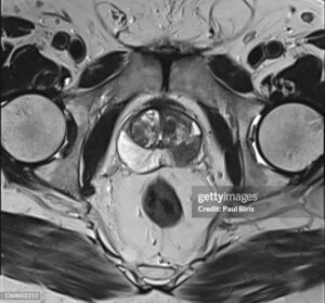
How prostate cancer occurs?
Prostate cancer, like most cancers, develops due to a complex interplay of factors, but here’s a breakdown of the key steps:
-
Genetic Mutations: In healthy cells, our DNA controls cell growth and division. In prostate cancer, specific mutations in the DNA of prostate gland cells lead to uncontrolled growth. These mutations can be inherited (though this is less common) or acquired over time due to various factors.
-
Abnormal Cell Growth: Cells with these mutations start growing and dividing more rapidly than normal cells. This abnormal growth can form a lump or mass in the prostate gland.
-
Tumor Formation: As these abnormal cells continue to multiply, they can clump together and form a tumor. It’s important to note that not all tumors are cancerous. Some prostate tumors are slow-growing and may never cause any problems (benign tumors).
-
Progression (not always the case): In some cases, these abnormal cells can develop further mutations that allow them to invade surrounding tissues and even spread to other parts of the body (malignant tumors). This is what defines prostate cancer.
Here are some additional factors that can contribute to the development of prostate cancer:
- Androgens: These are male sex hormones, including testosterone. Testosterone can stimulate the growth of both normal and abnormal prostate cells.
- Inflammation: Chronic inflammation in the prostate may play a role in the development and progression of prostate cancer, although the exact link is still being studied.
- Environmental factors: Exposure to certain chemicals or environmental toxins may be a potential risk factor, but more research is needed to understand the specific link.
It’s important to remember:
- The exact causes of prostate cancer are not fully understood.
- The development of prostate cancer is often a slow process that can take years, even decades.
- Early detection is crucial for successful treatment. Regular checkups and screenings with your doctor are important for prostate health.
If you have any further questions about prostate cancer or your individual risk factors, talking to your doctor is the best course of action.
How prostate cancer is detected ?
Early detection is key to successful treatment of prostate cancer. Here’s a breakdown of the most common methods used to detect prostate cancer:
Digital Rectal Exam (DRE):
- During a DRE, a doctor inserts a gloved, lubricated finger into the rectum to feel the prostate gland for any abnormalities like lumps, swelling, or changes in texture. This can be an indicator of cancer or other prostate conditions.
Prostate-Specific Antigen (PSA) Test:
- This is a blood test that measures the level of prostate-specific antigen (PSA) in the bloodstream. The prostate gland produces PSA, and elevated PSA levels can be a sign of prostate cancer, but it can also be caused by other factors like infection, inflammation, or benign enlargement of the prostate. A high PSA test result doesn’t necessarily mean cancer, and further tests will likely be recommended by a doctor.
**Here are some additional tests that may be used depending on the results of the initial screening and a doctor’s recommendation:
- Biopsy: If a DRE or PSA test suggests abnormalities, a doctor may recommend a biopsy. This involves taking a small tissue sample from the prostate using a thin needle for examination under a microscope to determine if cancer cells are present.
- Imaging tests: Imaging tests like ultrasound, MRI scans, or CT scans may be used to get a better view of the prostate and surrounding tissues. These can help determine the location and extent of potential cancer if a biopsy confirms its presence.
It’s important to remember:
- These are general guidelines, and a doctor will determine the most appropriate screening approach based on your individual risk factors and age.
- Not everyone with elevated PSA levels has prostate cancer, and some men with prostate cancer may have normal PSA levels.
- Early detection is crucial, so don’t hesitate to talk to your doctor about any concerns or to schedule regular checkups and screenings as recommended.
Here are some additional resources you might find helpful:
- National Cancer Institute – Prostate Cancer: [National Cancer Institute (.gov) prostate cancer ON National Cancer Institute (.gov) cancer.gov]
- American Cancer Society – Can Prostate Cancer Be Found Early?: [Can Prostate Cancer Be Found Early? | How to Detect Prostate Cancer? | American Cancer Society cancer.org]
How prostate cancer is caused?
The exact cause of prostate cancer is still not fully understood by scientists. However, research has identified several factors that can increase a man’s risk of developing the disease. These factors likely play a role in the abnormal cell growth that characterizes cancer:
- Genetics: Having a father, brother, or son diagnosed with prostate cancer increases your risk. Genes you inherit can influence your susceptibility to developing the disease.
- Age: The risk of prostate cancer increases significantly with age. Most cases are diagnosed in men over 50. Age-related changes in hormones and cell growth patterns are likely contributors.
- Hormones: Testosterone, the main male sex hormone, plays a role in prostate cell growth. Abnormalities in testosterone levels or how cells respond to testosterone might be involved in cancer development.
- Diet: A diet high in red meat and processed meats may be linked to an increased risk of prostate cancer. Conversely, a diet rich in fruits, vegetables, and whole grains might offer some protective benefits. The exact reasons behind these dietary associations are still being investigated.
- Obesity: Being overweight or obese can increase your risk of prostate cancer. Obesity can affect hormone levels and potentially contribute to abnormal cell growth.
Here are some additional details on how these factors might be involved:
- Mutations in genes: Certain genetic mutations can affect how cells grow and divide. These mutations may contribute to the uncontrolled cell growth seen in cancer.
- Changes in DNA: Over time, damage to a cell’s DNA can accumulate. This damage can lead to abnormal cell behavior and potentially cancer development.
- Chronic inflammation: Chronic inflammation in the prostate gland might create an environment that promotes abnormal cell growth.
How prostate cancer spreads ?
Prostate cancer can spread from the original tumor in the prostate gland to other parts of the body. Here’s a breakdown of the process:
Breakaway Cells: Cancerous cells within the prostate tumor may sometimes break away from the main mass. These breakaway cells can enter the bloodstream or lymphatic system (a network of vessels that drain fluid and waste from tissues).
Travel: Once in the bloodstream or lymphatic system, these cancerous cells can travel throughout the body.
Distant Sites: The cancer cells can get lodged in other organs or tissues where they can start to grow and form new tumors. These new tumors are called metastases (singular: metastasis).
Common Spread Sites: The most common places for prostate cancer to spread to are:
- Lymph nodes: These are small, bean-shaped organs located throughout the body that help filter out waste and infection. Lymph nodes near the prostate are the first potential targets for cancer spread.
- Bones: The bones, particularly the pelvis, spine, and ribs, are another frequent site for prostate cancer metastasis. Bone metastases can cause pain, weakness, and fractures.
- Other organs: In some cases, prostate cancer can spread to other organs like the lungs or liver, although this is less common.
Factors Affecting Spread: The likelihood and speed of prostate cancer spread depend on several factors, including:
- Grade of the cancer: Higher-grade cancers, which are more aggressive, are more likely to spread than lower-grade cancers.
- Stage of the cancer: The stage refers to the extent of cancer spread. Early-stage cancers are confined to the prostate, while later stages involve lymph node or distant organ involvement.
- Patient’s health: A person’s overall health and immune system function can influence the spread of cancer cells.
If you have prostate cancer, your doctor will consider these factors to assess your risk of spread and determine the most appropriate treatment course. Early detection and treatment can significantly improve the chances of controlling prostate cancer and preventing its spread.
How prostate cancer test is done? How prostate cancer diagnosed?
How many radiation treatments for prostate cancer?
The number of radiation treatments you’ll receive for prostate cancer depends on several factors, including:
- Stage and grade of your cancer: More advanced or aggressive cancers may require a higher total dose of radiation, translating to more treatment sessions.
- Type of radiation therapy: Different radiation therapy techniques deliver varying radiation doses per session.
- Your overall health and tolerance for treatment: Doctors consider your individual health to determine the most appropriate treatment schedule.
Here’s a general breakdown of the number of radiation treatments for prostate cancer:
-
Conventional Radiation Therapy: This is the most common type, typically involving daily treatments (Monday through Friday) for a total of:
- 35-45 treatments: This is the usual range for curative treatment of prostate cancer.
-
Hypofractionation: This approach uses higher radiation doses per session but fewer total sessions. It might be an option for certain patients. The number of sessions can vary but might be:
- 20-28 treatments: This is a possible range for hypofractionated radiation therapy.